Optometry 360
  • Home
  • Conferences
  • Videos
  • About Us
    • About Us – Mission
    • Media Partners
    • Advisory Board
    • Industry Council
    • Business Team

What are you looking for?

  • Conference Roundup
  • Contact Lenses
  • Diagnostics
  • Glaucoma
  • Myopia
  • Neurotrophic Keratitis
  • Ocular Surface Disease
  • Pediatrics
  • Practice Management
  • Presbyopia
  • Retina
  • Spotlight Series
The evolving role of the optometrist in nonsurgical cataract management
Optometry 360
  • Home
  • Conferences
  • Videos
  • About Us
    • About Us – Mission
    • Media Partners
    • Advisory Board
    • Industry Council
    • Business Team
Home > Clinical examination findings help predict meibomian gland atrophy, study finds
  • Ocular Surface Disease

Clinical examination findings help predict meibomian gland atrophy, study finds

Kelsey Moroz

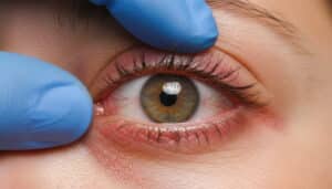
Gently pressing on the meibomian glands to check how easily they release oil is a strong way to predict meibomian gland atrophy (MGA), according to a study. Although other factors like lipid layer thickness and vascular engorgement also contribute, clinical examination alone does not fully explain MGA variability.

Researchers evaluated dry eye symptoms using the SPEED questionnaire and examined various clinical parameters, including lipid layer thickness, Schirmer I scores, lid margin characteristics, and meibomian gland secretion factors. Infrared meibography assessed MGA severity, while multivariate binary logistic regression identified key predictors.

Expressibility of meibomian gland secretions was the strongest predictor of MGA severity, with an odds ratio of 2.604 (P = 0.020), sensitivity of 85.7%, and specificity of 59.1%. The Foulks-Bron (FB) score was also significantly correlated with MGA severity (R = 0.372, P = 0.014). Lipid layer thickness (P = 0.042) and vascular engorgement (P = 0.032) were found to be predictive.

Reference
Suraneni S, Mulpuri L, Habib M, et al. Efficacy and utility of clinical examination in predicting meibomian gland atrophy. Cont Lens Anterior Eye. 2025;102405. doi: 10.1016/j.clae.2025.102405. Epub ahead of print. PMID: 40090800.

Share

Related Content

  • Ocular Surface Disease

Smartphone reminders improve short-term artificial tear adherence in dry eye disease

  • Ocular Surface Disease

Childhood-onset allergic eye disease linked to greater ocular morbidity

  • Ocular Surface Disease

Perceived stress may precede worsening dry eye symptoms in young adults

  • Ocular Surface Disease

Post-menopausal women show more severe evaporative dry eye disease

  • Ocular Surface Disease

Dry eye diagnostic practices remain inconsistent worldwide, survey finds

  • Ocular Surface Disease

Spotlight on VEVYE®

Share

Advisory Board

Kaleb Abbott, OD, MS, FAAO

Clark Chang, OD

Jessica Haynes, OD, FAAO, FORS, Dipl ABO

Monica Kalia, OD

Cecelia Koetting, OD, FAAO, DipABO

Roya Attar, OD, MBA, DHA, FAAO

Hardeep Kataria, OD, FAAO

Marc R. Bloomenstein, OD, FAAO

Elise Kramer, OD

See All
Optometry 360

Optometry 360® is a dynamic digital platform dedicated to advancing the field of eye care.

Get to Know Us

  • Home
  • About Us
  • Media Partners
  • Advertising Policy
  • Our Advisory Board

Sign up for our Newsletter

Sign up for our Newsletter to get our newest articles instantly!

  • Privacy Policy
  • Advertising Policy
  • Medical Disclaimer
IHM Logo

© 2026 Optometry 360 is a trademark of International Healthcare Media, LLC. All rights Reserved

  • MedJournal360 Icon
  • RareDisease360 Icon
  • MyHero360 Icon
  • Optometry360 Icon
  • Ophthalmology360 Icon